Innovative Insights on Finger Tendon Health
The Crucial Role of Finger Tendons in Daily Function

Understanding Finger Tendons
Finger tendons are essential components of the hand, allowing for precise and powerful movements. These intricate structures connect the muscles in the forearm to the bones in the fingers, enabling actions such as grasping, pinching, and typing. Understanding their anatomy and function is crucial for recognizing and addressing potential issues. These delicate tissues are responsible for a wide range of activities, from the simple act of picking up a pen to the more complex motions involved in playing a musical instrument.
The tendons within the hand are organized in a complex network, facilitating a wide range of motions. Proper function relies on the smooth gliding of the tendons within their sheaths. Damage or inflammation to these tendons can lead to significant limitations in hand function. Early diagnosis and appropriate treatment are key to minimizing long-term problems.
The Anatomy of Finger Tendons
The intricate anatomy of finger tendons involves multiple components working in harmony. These include the flexor tendons, responsible for bending the fingers, and the extensor tendons, responsible for straightening them. Each tendon is encased in a protective sheath, which allows for smooth movement and reduces friction. This intricate system permits a wide range of precise and powerful hand movements.
These tendons are not simply passive structures. They are active participants in the complex dance of hand movement. They are tightly bound to the bones and ligaments, ensuring precise control and stability. This intricate interplay of components is vital for the delicate movements required for tasks ranging from writing to playing musical instruments.
Maintaining Healthy Finger Tendons
Maintaining the health of finger tendons is crucial for overall hand function. Regular hand exercises, proper posture during activities that involve repetitive hand movements, and avoiding excessive strain are important preventive measures. A balanced diet rich in nutrients that support healthy tissue repair can further contribute to overall tendon health. This proactive approach can help prevent the development of problems like tendonitis or other conditions.
Keeping your hands warm and protecting them from extreme temperatures is also important. This is particularly important in cold climates or when engaging in activities that can expose your hands to cold conditions. Proper hand care can significantly reduce the risk of developing tendon-related issues. Maintaining optimal hand health often involves a combination of habits and practices.
Common Issues Affecting Finger Tendons
Several conditions can affect the health and function of finger tendons. Tendonitis, a common ailment, results from inflammation of the tendons, often due to repetitive strain. Other issues include tendon ruptures, which can occur due to sudden forceful movements. These injuries can significantly limit hand function. Prompt diagnosis and appropriate treatment are essential to managing these issues.
Carpal tunnel syndrome, although not directly affecting the tendons themselves, can cause significant compression of the tendons and nerves in the wrist, leading to pain and numbness in the hands and fingers. Therefore, recognizing the signs and symptoms of these conditions is crucial for early intervention.
Innovative Diagnostic Techniques for Early Detection
Advanced Imaging Technologies
Modern imaging techniques like ultrasound, magnetic resonance imaging (MRI), and computed tomography (CT) scans are revolutionizing the field of diagnostics. These methods allow for non-invasive visualization of internal structures, enabling the detection of subtle abnormalities that might otherwise go unnoticed. High-resolution imaging capabilities are crucial in identifying early stages of disease, often before symptoms manifest, significantly improving treatment outcomes. The ability to visualize intricate anatomical details and physiological processes in real-time provides invaluable information for clinicians, facilitating precise diagnoses and tailored treatment plans. This advancement in technology is particularly beneficial in cases of suspected early-stage finger conditions, allowing for prompt intervention and minimizing potential long-term complications.
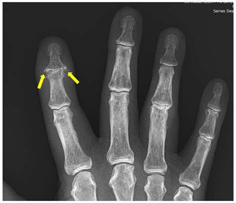
Furthermore, advancements in contrast agents and imaging protocols are enhancing the specificity and sensitivity of these techniques. These enhancements allow for a more detailed and precise evaluation of the affected area, leading to earlier and more accurate diagnoses. The development of specialized imaging protocols tailored specifically to finger structures is crucial for accurate assessment of potential pathologies, facilitating the identification of early signs of conditions like arthritis or fractures, which can have profound implications for the patient's overall health and well-being.
Molecular Diagnostics and Biomarkers
The application of molecular diagnostics, focusing on the analysis of genetic material and proteins, is emerging as a powerful tool for early detection. By identifying specific biomarkers associated with diseases, clinicians can detect subtle changes in the body long before the onset of noticeable symptoms. This approach has the potential to identify individuals at risk of developing certain conditions, enabling preventive measures and interventions before disease progression.
Analyzing genetic mutations and protein expression patterns in tissue samples obtained from the finger can offer valuable insights. This approach can differentiate between various conditions with similar symptoms, leading to more accurate diagnoses. The ability to identify specific molecular markers associated with early-stage conditions significantly contributes to personalized medicine approaches, allowing for targeted therapies and optimized treatment strategies, ultimately improving patient outcomes.
The development of highly sensitive and specific molecular diagnostic tests is critical for early detection of various finger conditions. These tests can analyze minute quantities of biological material, offering unparalleled precision in identifying early disease processes. This precision is particularly valuable in finger conditions where early intervention is crucial for preventing long-term damage and maintaining optimal functionality.
Advanced molecular techniques are poised to revolutionize the diagnosis of finger conditions. Early detection through these methods will lead to improved patient outcomes and contribute significantly to the field of hand surgery and orthopedics.
The use of genetic sequencing and proteomics can help identify specific mutations or protein abnormalities linked to particular finger conditions. This can potentially lead to earlier intervention and potentially prevent the progression of the disease.
Emerging Therapeutic Strategies for Tendon Repair and Regeneration
Stem Cell Therapies for Tendon Repair
Stem cell therapies represent a promising frontier in tendon repair and regeneration. These therapies leverage the inherent regenerative capacity of stem cells, either derived from the patient's own body (autologous) or from other sources (allogeneic or xenogeneic). Autologous stem cells, such as mesenchymal stem cells (MSCs) and adipose-derived stem cells (ADSCs), have shown promising results in preclinical models, demonstrating the potential to promote tendon healing by stimulating collagen synthesis, modulating inflammation, and reducing scar tissue formation. Further research is crucial to optimize these approaches, including the precise delivery methods and the selection of appropriate stem cell sources.
Challenges remain in translating these promising preclinical findings into effective clinical treatments, including ensuring sufficient stem cell delivery to the injured tendon, minimizing the risk of immune rejection in allogeneic or xenogeneic approaches, and optimizing the therapeutic environment to support stem cell differentiation and function. The future likely holds more sophisticated strategies for stem cell delivery, such as using biocompatible scaffolds or targeted drug delivery systems.
Biomaterials and Scaffolds for Tendon Regeneration
Biomaterials and scaffolds play a critical role in tendon regeneration by providing a supportive structure for the healing process. These materials are designed to mimic the natural extracellular matrix (ECM) of tendons, encouraging cell adhesion, proliferation, and differentiation. Various biomaterials, including synthetic polymers and natural polymers like collagen and chitosan, are being investigated. These materials can be formulated into different shapes and structures to precisely guide tissue regeneration.
The key to successful biomaterial application lies in creating a scaffold that is biocompatible, bioresorbable, and promotes the formation of a robust and functional tendon structure. The scaffold should also facilitate the integration of cells and growth factors, further encouraging tendon regeneration.
Growth Factors and Cytokines in Tendon Healing
Growth factors and cytokines are natural mediators of cellular processes, and their strategic application can significantly influence tendon repair and regeneration. These factors can promote cell proliferation, differentiation, and migration, stimulating collagen synthesis and matrix deposition. Examples include transforming growth factor-β (TGF-β), platelet-derived growth factor (PDGF), and vascular endothelial growth factor (VEGF). Precisely controlling the release and delivery of these factors is crucial for optimal therapeutic outcomes.
Careful consideration must be given to the optimal concentration, duration, and delivery method of these factors to avoid unwanted side effects. Further research is needed to refine the use of these factors, potentially through combining multiple growth factors or using gene therapy approaches to enhance their local production.
Gene Therapy Approaches for Enhanced Tendon Repair
Gene therapy holds immense potential for enhancing tendon repair and regeneration by directly influencing the expression of genes involved in tissue healing. Modifying the genetic makeup of cells to overexpress key proteins or downregulate factors that hinder repair can create a more favorable environment for tendon regeneration. For example, gene therapy targeting specific signaling pathways could potentially promote collagen synthesis and reduce scar tissue formation.
Combination Therapies for Synergistic Effects
Combining different therapeutic strategies, such as stem cell therapies, biomaterials, growth factors, and gene therapy, may offer synergistic effects, leading to more effective tendon repair and regeneration. This approach could enhance the regenerative capacity of the injured tendon by addressing multiple aspects of the healing process simultaneously. For example, a combination of a specific growth factor, a biocompatible scaffold, and stem cell therapy could potentially create an optimal microenvironment for enhanced tissue regeneration.
Novel Therapeutic Techniques and Future Directions
Innovative techniques such as ultrasound-guided stem cell injection, targeted drug delivery systems, and 3D bioprinting are paving the way for more precise and effective tendon repair and regeneration strategies. These approaches can improve the delivery and localization of therapeutic agents, thus minimizing unwanted side effects and enhancing the effectiveness of treatment. Further research into these advanced techniques will likely lead to even more personalized and targeted approaches in the future.


