Advanced Diagnostics in Hand Bone Health
Radiographic Techniques for Hand Assessment
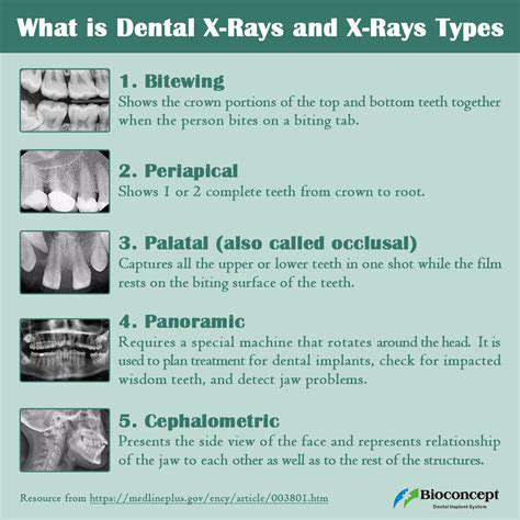
Radiographic imaging, a cornerstone of hand bone assessment, employs various techniques to visualize the intricate structures of the hand. Different projections, such as posteroanterior (PA) and lateral views, are crucial for capturing complete anatomical details. Understanding the appropriate positioning and technique is vital for obtaining high-quality images, minimizing artifacts, and ensuring accurate diagnoses. Radiographers must meticulously follow protocols to ensure consistency and reproducibility in image acquisition.
Furthermore, specialized views, such as oblique projections, may be necessary to assess specific joints or bone segments in greater detail. These specialized techniques allow for a more comprehensive evaluation of the hand's anatomy and can be particularly important in cases of suspected fractures or joint dislocations. Understanding the rationale behind each technique is essential for radiologists and clinicians interpreting the images.
Importance of Image Quality in Hand Radiography
Achieving optimal image quality is paramount in hand radiography. Factors such as patient positioning, exposure factors, and image processing significantly influence the diagnostic accuracy of the images. Proper patient positioning ensures that the structures of interest are visualized without overlapping or obscuring details. Accurate exposure factors are essential to avoid overexposure or underexposure, both of which can compromise image quality and diagnostic interpretation.
Bone Morphology and Radiographic Appearance
Radiographic assessment of hand bones relies heavily on the recognition of normal bone morphology. The shape, size, and density of the bones provide valuable clues about potential abnormalities. Radiographers and clinicians must have a thorough understanding of the normal radiographic appearance of each bone in the hand, including the carpals, metacarpals, and phalanges. Variations in bone morphology can be subtle but significant, indicating underlying conditions or developmental issues.
Fractures and Dislocations: Radiographic Detection
Radiographic imaging is the primary modality for detecting fractures and dislocations in the hand. The presence of a fracture line or displacement of bone segments is often readily apparent on radiographs. Different types of fractures, such as transverse, oblique, or comminuted fractures, exhibit characteristic appearances on radiographs. A thorough understanding of fracture patterns is critical for accurate diagnosis and appropriate treatment planning.
Joint Spaces and Articulations: Evaluation of Carpal and Metacarpophalangeal Joints
Radiographic evaluation of joint spaces and articulations is crucial for assessing the health of the hand's joints. The width and clarity of joint spaces are key indicators of joint health. Narrowing or widening of joint spaces can suggest various pathologies, including arthritis, inflammation, or trauma. Careful analysis of the carpal and metacarpophalangeal joints is essential for comprehensive assessment.
Soft Tissue Assessment and its Relation to Bone
While the primary focus is on bone, radiographic images also provide valuable information about the surrounding soft tissues. Soft tissue swelling, edema, or masses can be identified and correlated with potential bone pathology. The relationship between soft tissue and bone abnormalities is important in understanding the underlying cause of symptoms. Radiologists need to carefully evaluate the soft tissue structures alongside the bony structures to get a complete picture.
Case Studies and Clinical Correlations
The application of radiographic imaging in hand bone assessment is best understood through real-world case studies. By analyzing various cases, including those involving fractures, dislocations, arthritis, and tumors, radiologists can develop a deeper understanding of the diagnostic capabilities of radiographic imaging. Clinical correlations between radiographic findings and patient symptoms are essential for accurate diagnosis and appropriate treatment.
