Next Level Insights into Wrist Joint Mechanics
The Interplay of Bones and Cartilage
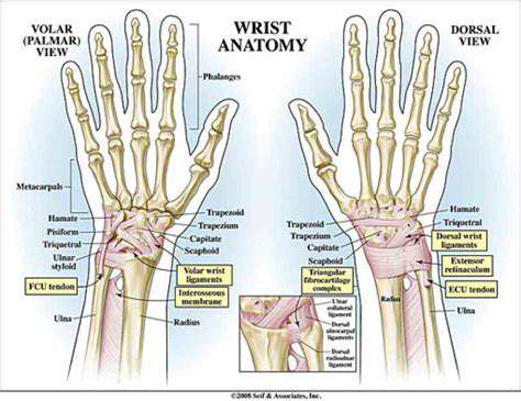
The wrist, though seemingly simple, is a marvel of anatomical engineering. A complex arrangement of eight small carpal bones, meticulously positioned and held together by ligaments, forms the foundation of this intricate joint. These bones, acting in concert with the radius and ulna from the forearm, provide a remarkable range of motion, crucial for everyday tasks like grasping, writing, and typing. The interplay of these bones is also critical for shock absorption, allowing us to handle impacts and forces without immediate injury.
Cartilage, a smooth, resilient tissue, plays a vital role in reducing friction between the bones during movement. This smooth surface allows for seamless gliding and minimizes wear and tear on the joint. The delicate balance between the structure of the bones and the cushioning provided by cartilage is essential for maintaining wrist health and function over a lifetime. Without this intricate arrangement, simple actions would be painful and potentially damaging.
Ligaments and Tendons: The Supporting Cast
The wrist's stability relies heavily on a network of strong ligaments. These fibrous bands connect the bones, providing crucial support and preventing excessive movement. Ligaments are vital for maintaining the wrist's structural integrity, ensuring that the bones remain in their proper alignment during various activities, from lifting objects to engaging in sports. Damage to these ligaments can lead to instability and significant pain, hindering daily tasks and requiring careful medical attention.
Tendons, another crucial component, connect muscles to bones. These strong cords transmit the force generated by muscles to the bones, enabling us to perform actions like flexing and extending the wrist. Damage to tendons, often resulting from overuse or injury, can cause significant pain, swelling, and limitations in wrist function. Proper care and attention to tendon health are essential for maintaining optimal wrist performance.
Nerves: The Crucial Communication Network
The intricate network of nerves within the wrist plays a vital role in transmitting signals between the brain and the muscles. These signals allow us to consciously control our wrist movements, enabling complex tasks like writing, playing musical instruments, and performing intricate hand movements. Damage to these nerves can result in numbness, tingling, or pain in the hand and wrist, as well as a loss of sensation and motor control. Understanding the pathways and functions of these nerves is critical for diagnosis and treatment of various wrist conditions.
Muscles: The Driving Force Behind Movement
The muscles surrounding the wrist are essential for a wide range of movements, from simple wrist flexion and extension to more complex tasks. These muscles, working in coordinated patterns, enable us to perform the precise and varied movements required for daily tasks and activities. Understanding the intricacies of muscle function and activation is vital for restoring optimal wrist function after injury or surgery.
Blood Supply: The Lifeblood of the Wrist
Adequate blood supply is crucial for providing nutrients and oxygen to the tissues of the wrist. Blood vessels, branching throughout the area, ensure that all components of the wrist receive the necessary resources to function properly. Compromised blood supply can lead to tissue damage and potentially serious complications. Maintaining a healthy circulation to the wrist is essential for preventing issues and promoting healing in case of injury.
Exploring the Role of Carpal Bones in Wrist Mechanics
Understanding the Carpal Bones
The carpal bones, a collection of eight small bones, form the wrist joint. These intricate components are crucial for the complex mechanics of hand movement, allowing us to perform a wide range of tasks from gripping objects to delicate manipulations. Their precise arrangement and articulation with other bones are fundamental to the wrist's flexibility and strength.
Understanding the individual carpal bones is key to comprehending the wrist's function as a whole. Each bone plays a specific role in the overall structure and movement of the wrist, and their coordinated actions determine the range of motion possible.
The Anatomy of the Carpal Bones
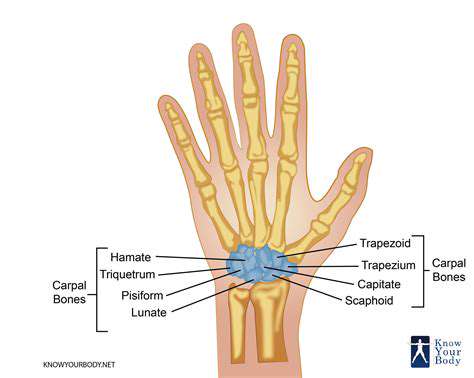
The carpal bones are arranged in two rows, proximal and distal. This arrangement allows for a varied range of motion, enabling the wrist to bend, extend, and rotate. The proximal row, comprised of the scaphoid, lunate, triquetrum, and pisiform bones, articulates with the radius and ulna bones of the forearm.
The distal row, consisting of the trapezium, trapezoid, capitate, and hamate bones, are connected to the metacarpal bones, forming the foundation of the hand.
The Scaphoid: A Crucial Bone
The scaphoid, located in the proximal row, is one of the most frequently fractured carpal bones due to its prominent position and the forces it bears during wrist movements. It is a crucial component of the wrist's stability and plays a significant role in the wrist's ability to support weight and handle stress. Its articulate design connects the radius to the middle section of the wrist.
The Lunate: A Key Player
The lunate, also part of the proximal row, is a crescent-shaped bone that forms a critical link between the radius and the other carpal bones. It's vital in maintaining the wrist's alignment and stability during various movements. A critical component of the wrist's intricate system.
The Role in Hand Movement
The carpal bones, working in concert, enable a wide range of hand movements. These movements include flexion, extension, and abduction, allowing us to perform tasks ranging from gripping objects to writing and typing. The complex interplay of these bones allows for the dexterity and precision required in everyday activities.
The smooth articulation of the carpal bones is essential for the wrist's mobility and overall functionality. These bones are not just static structures; their movements are intrinsically linked to the functionality of the entire hand.
Clinical Significance and Disorders
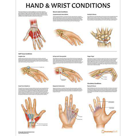
Conditions like carpal tunnel syndrome, fractures, and arthritis can significantly impact the carpal bones and their functionality. Diagnosing and treating these conditions require a thorough understanding of the carpal bones' anatomy and their role in the wrist's movement. This understanding allows for effective treatments that restore proper function and alleviate pain.
Identifying and addressing these issues promptly is crucial to prevent long-term complications and maintain the integrity of the wrist joint. Early detection and appropriate treatment strategies are essential to preserving the wrist's overall health and movement.
Maintaining Carpal Bone Health
Maintaining healthy carpal bones is essential for overall hand and wrist health. Practicing proper ergonomics in daily activities, maintaining a healthy weight, and engaging in regular exercise that strengthens the wrist and forearm muscles are all important. A balanced diet that supplies essential nutrients also contributes to the health of the carpal bones.
Regular hand stretches and exercises can contribute to improved wrist flexibility and strength, reducing the risk of injury. Protecting the wrists from repetitive stress and impact is also crucial in preventing carpal bone disorders. These are just a few of the steps that can be taken to maintain healthy carpal bones.
Dissecting the Ligamentous Support System: Maintaining Wrist Stability
Understanding the Crucial Role of Wrist Ligaments
Wrist stability is paramount for a wide array of daily activities, from simple tasks like picking up objects to more complex movements like playing musical instruments or participating in sports. The intricate network of ligaments within the wrist acts as a crucial support system, preventing excessive movement and ensuring the delicate bones of the wrist remain aligned. These ligaments, essentially strong bands of connective tissue, are strategically positioned to limit undesirable movements, thereby safeguarding the wrist from potential injuries and maintaining its optimal function.
Comprehending the anatomical arrangement of these ligaments—their precise attachments, and the forces they endure—is essential for grasping their significance. Understanding the intricate interplay between the ligaments, tendons, and muscles surrounding the wrist is key to appreciating the comprehensive nature of wrist stability. This knowledge is instrumental in developing effective strategies for injury prevention and rehabilitation, thereby promoting optimal wrist health and function.
Identifying the Key Ligaments and Their Functions
Several key ligaments contribute to the overall stability of the wrist. The scapholunate ligament, for instance, plays a critical role in maintaining the proper alignment between the scaphoid and lunate bones, preventing excessive sideways displacement. The lunotriquetral ligament similarly safeguards the alignment of the lunate and triquetral bones, ensuring stability during wrist movements. These ligaments, along with the radial and ulnar collateral ligaments, work in concert to provide a robust, multi-directional support system for the wrist joint.
The importance of these ligaments extends beyond simple support. They function as critical stabilizers, preventing excessive flexion, extension, and deviation of the wrist. Their ability to withstand substantial forces without yielding is paramount for preventing injuries, particularly during activities involving repetitive movements or high impact.
Maintaining Wrist Ligament Health: A Proactive Approach
Maintaining the health of wrist ligaments is crucial for preventing injuries and ensuring long-term wrist function. A proactive approach, incorporating appropriate warm-up exercises, proper technique during activities, and the use of supportive gear when necessary, can significantly reduce the risk of ligament damage. This proactive approach is vital for athletes and individuals performing repetitive wrist movements, as it helps prevent overuse injuries and promote optimal ligament health.
Furthermore, maintaining a healthy weight and engaging in regular strength training exercises that target the muscles surrounding the wrist can contribute to improved wrist stability and reduce stress on the ligaments. A balanced diet rich in nutrients essential for connective tissue health, such as vitamin C and collagen-boosting foods, further supports the resilience of wrist ligaments. Regular physical therapy, especially after injuries, is also essential for restoring and strengthening these crucial structures.
The Muscle Contributions to Wrist Dynamics: Beyond the Surface
Muscle Actions in Wrist Extension
The primary muscles responsible for wrist extension are the extensor carpi radialis longus and brevis, and the extensor carpi ulnaris. These muscles, originating from the lateral epicondyle of the humerus and other points along the posterior forearm, contract to produce wrist extension. Understanding the precise actions of these muscles is crucial for rehabilitation and therapeutic exercise design. This movement is essential for tasks like lifting objects and maintaining a stable grip.
The extensor carpi radialis longus primarily extends and abducts the wrist, while the extensor carpi radialis brevis primarily extends and abducts the wrist. The extensor carpi ulnaris extends and adducts the wrist. This intricate interplay of muscle action allows for a wide range of wrist movements, contributing to overall hand function.
Muscle Contributions to Wrist Flexion
Wrist flexion is primarily controlled by the flexor carpi radialis, flexor carpi ulnaris, and palmaris longus. These muscles originate from the medial epicondyle of the humerus and the anterior forearm. The synergistic action of these muscles ensures smooth and controlled wrist flexion. This is critical for gripping objects and performing fine motor tasks.
The flexor carpi radialis flexes and abducts the wrist. The flexor carpi ulnaris flexes and adducts the wrist. The palmaris longus primarily flexes the wrist. Understanding these actions is vital for diagnosing and treating wrist injuries.
The Role of Intrinsic Muscles
Intrinsic muscles of the hand, such as the interossei and lumbricals, play a vital role in wrist movement, though not as primary movers. These muscles are located within the hand and contribute to fine motor control and precise movements of the fingers and thumb. Their subtle but essential contributions to wrist function are often overlooked. Understanding their role is particularly important in situations requiring dexterity and precision.
The Importance of Wrist Stability
Maintaining wrist stability is crucial for preventing injuries and ensuring optimal function. The coordinated action of multiple wrist muscles, including the aforementioned extensors and flexors, contribute to this stability. These muscles work in concert to stabilize the wrist joint during various activities. This stability is essential for preventing injury during activities such as gripping and lifting.
Neuromuscular Control and Coordination
The intricate interplay between the nervous system and the muscles of the wrist is essential for smooth and controlled movements. Proper neuromuscular control ensures coordinated contractions and relaxation of muscles, enabling precise wrist movements required for daily activities. Neuromuscular control is essential for performing tasks ranging from writing to playing musical instruments. Dysfunction in this area can lead to weakness, instability, and pain.
Muscle Imbalances and Their Impact
Muscle imbalances, where one group of wrist muscles is significantly stronger than another, can lead to wrist pain and injury. Overuse or repetitive strain on a specific set of muscles can cause imbalances that lead to tendinopathy, carpal tunnel syndrome, or other conditions. Identifying and correcting muscle imbalances is a critical step in preventing and treating wrist injuries. Physical therapy and targeted exercises can help restore balance.
Clinical Significance of Muscle Actions
Understanding the specific actions and interactions of wrist muscles is crucial for clinicians in diagnosing and treating wrist injuries. The ability to isolate and assess the strength and range of motion of individual muscles allows for a more accurate assessment of the extent of the injury and the development of effective rehabilitation strategies. Accurate diagnoses are essential for prescribing appropriate treatment. This includes recognizing the subtle variations in muscle function that might indicate underlying issues.
Advanced Techniques for Assessing Wrist Function: From Observation to Imaging
Observation-Based Assessments
Initial assessments of wrist function often begin with a careful observation of the patient's posture and movement patterns. A skilled clinician can glean valuable information about potential limitations or asymmetries by observing the patient's ability to perform everyday tasks like grasping objects, lifting weights, and turning doorknobs. Careful attention to subtle differences in range of motion, pain provocation, and overall ease of movement can provide crucial clues about the underlying cause of wrist dysfunction, particularly in cases where objective measures are inconclusive or not yet available.
Beyond simple observational assessments, the clinician should also document the patient's description of their symptoms, including the location, intensity, and duration of pain. This subjective information, coupled with the objective observations, provides a more holistic picture of the patient's experience and guides the subsequent diagnostic process. Careful documentation of these observations is essential for tracking progress over time and for communicating findings effectively to other healthcare professionals.
Manual Muscle Testing and Range of Motion
Manual muscle testing provides a quantifiable assessment of the strength and endurance of the wrist's intrinsic and extrinsic muscles. Specific tests are designed to isolate the function of individual muscle groups, allowing the clinician to identify potential muscle weakness or atrophy. This process involves applying resistance to the patient's active movements, assessing the strength and endurance of the muscles involved in wrist flexion, extension, radial deviation, and ulnar deviation. A detailed evaluation of the range of motion in each of these directions, both actively and passively, provides further insights into the joint's mobility and potential restrictions.
Accurate measurement of range of motion is crucial. Using standardized goniometry techniques, clinicians can precisely quantify the angles of flexion, extension, radial deviation, and ulnar deviation. These measurements are documented and compared to normative values to assess the degree of limitation or impairment. Such precise measurements provide objective data that can be used for comparison throughout the diagnostic and treatment process, helping to track improvements and monitor the effectiveness of interventions.
Imaging Techniques for Comprehensive Evaluation
While observation and manual testing are fundamental, imaging techniques offer a more detailed view of the wrist's internal structures. X-rays are invaluable for evaluating bone alignment, identifying fractures, and detecting signs of arthritis. Furthermore, advanced imaging modalities like MRI and CT scans provide a detailed visualization of the soft tissues, tendons, ligaments, and cartilage within the wrist. These techniques allow for the identification of tears, inflammation, and other pathologies that may not be apparent through physical examination alone.
MRI scans are particularly useful for assessing soft tissue structures, providing detailed images of tendons, ligaments, and nerves. This information can be crucial for diagnosing conditions like carpal tunnel syndrome, De Quervain's tenosynovitis, and other tendon-related injuries. Proper interpretation of these images is critical, often requiring consultation with a radiologist to ensure accurate diagnosis and appropriate treatment planning.
Read more about Next Level Insights into Wrist Joint Mechanics